
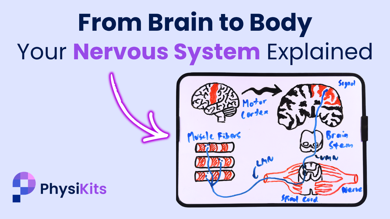
From Brain to Body: Your Nervous System Explained
Feb 27, 2026Did you know that some neurons in your spinal cord are over 1 metre long — making them the longest cells in your body?
And that the signal letting you reach out and pick something up travels from brain to muscle via just two?
In this post, we're breaking down how the nervous system actually works — how signals travel from brain to body and back again — so that you have a clear foundation for understanding what goes wrong in different neurological conditions.
This article is a companion to our PK Explains video on the same topic. If you prefer to watch rather than read, check out the full episode on YouTube!
The Humble Neuron
Every movement you've ever made, every sensation you've ever felt, comes down to electricity and chemistry moving through specialised cells called neurons (or nerve cells).
Unlike most cells in your body, neurons are built to transmit information.
They have three main parts.
- Dendrites receive signals from other neurons.
- The cell body houses the internal machinery of the cell.
- And the axon — a long, cable-like structure — carries signals onward, ending in axon terminals that form connections with other neurons at tiny gaps called synapses.
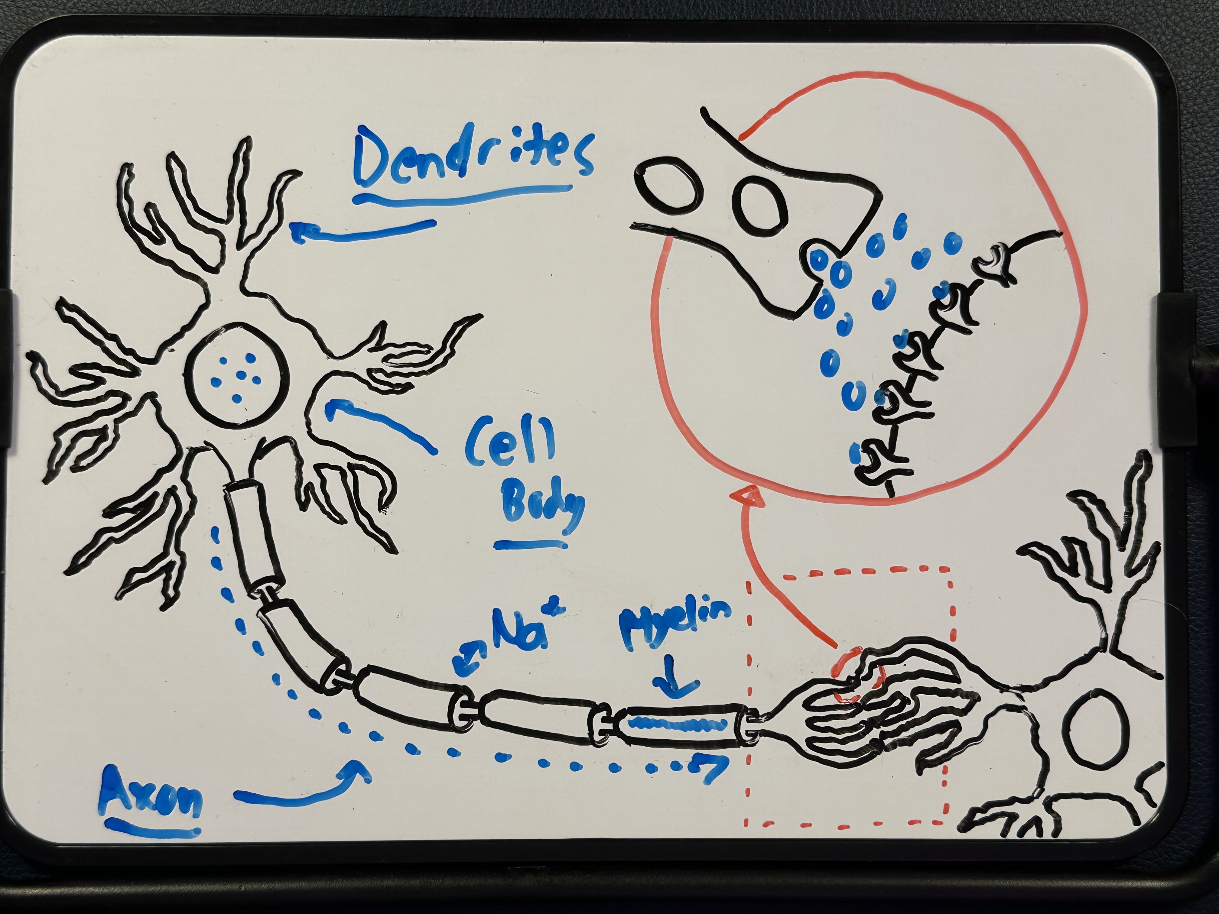
Here's the key idea: neurons communicate using a combination of electrical and chemical signals.
Inside the neuron, information travels as an action potential — a rapid shift of electrically charged ions (like sodium and potassium) across the cell membrane. That shift creates a small electrical impulse that propagates down the axon.
When that electrical signal reaches the end of the axon, it can't just jump to the next cell. Instead, it triggers the release of neurotransmitters — chemical messengers that cross the synapse and activate the next neuron.
Basically, electricity travels along the wire, chemicals cross the gap, then electricity continues again.
And just like in electrical wires, insulation is needed to keep that signal clear. The fastest axons are wrapped in a fatty layer called myelin, which protects the signal from interruption and allows it to travel quickly and efficiently.
Now while one neuron sounds complex on its own, remember that you have billions of them (commonly estimated at around 86 billion), all interconnecting in the vast network that is your nervous system.
Central vs Peripheral
Broadly speaking, the nervous system is divided into two major parts:
Central nervous system (CNS)
The CNS consists of your brain and spinal cord. The brain is the main control centre where signals are processed and generated. Different regions specialise in different functions — movement, sensation, memory, vision — but that complexity deserves its own dedicated video and blogpost in the future.
(Though we did touch on it in this episode of NeuroNotes!)
For now, a key concept to know is that the outermost layer of the brain, called the cortex, is where most signal processing and generation takes place. The cortex is dense with neuron cell bodies and dendrites, giving it a darker colour — which is where the term "grey matter" comes from.
Deeper structures, by contrast, are rich in myelinated axons carrying signals to and from the cortex, and their lighter colour gives them the name "white matter".
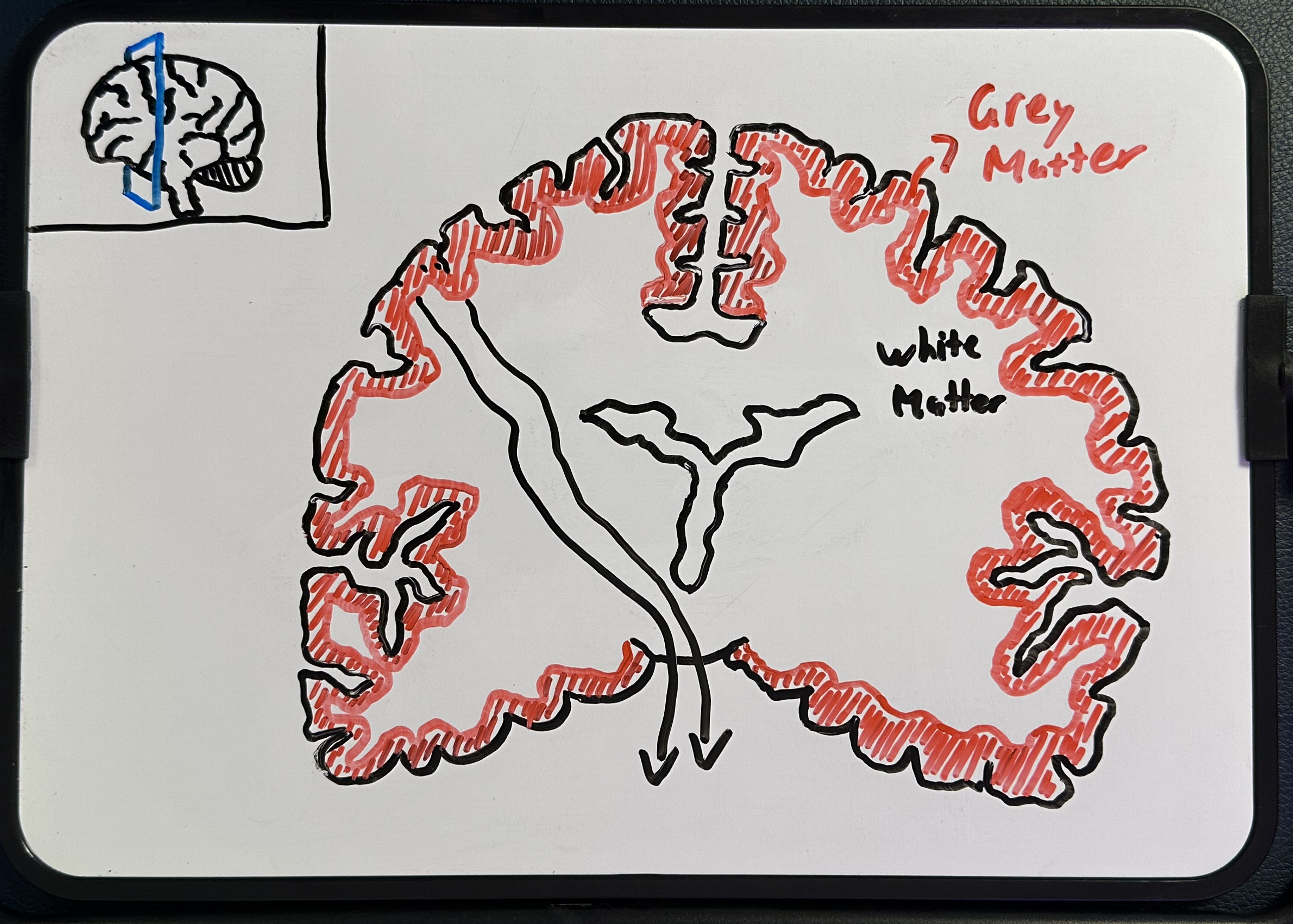
Signals pass through the brainstem to the spinal cord, which is packed with long axons acting as a highway for signals travelling to and from the body.
An important detail: At the brainstem junction, many motor and sensory pathways cross over from left to right. This is why the left side of your brain senses and controls the right side of your body, and vice versa!
Peripheral nervous system (PNS)
Below the spinal cord, we enter the territory of the peripheral nervous system — this includes all the nerves that branch out from the CNS into your body. While 12 cranial nerves branch directly from the brain, the majority of peripheral nerves exit from the spinal cord through structures called nerve roots, forming the peripheral nerves that travel to muscles, skin, joints, and organs.
From the moment a nerve exits the spinal cord to its final destination, that entire length is a bundle of individual nerve cells running in parallel. This is why some nerve axons — like those in the sciatic nerve running from your spine to your toes — can reach over 1 metre in length.
To recap: the brain and spinal cord form the central command centre, the peripheral nerves are the delivery network, and everything is connected by long, insulated cables called axons.
Motor Output: Brain to Muscle
Let's trace a signal through the system.
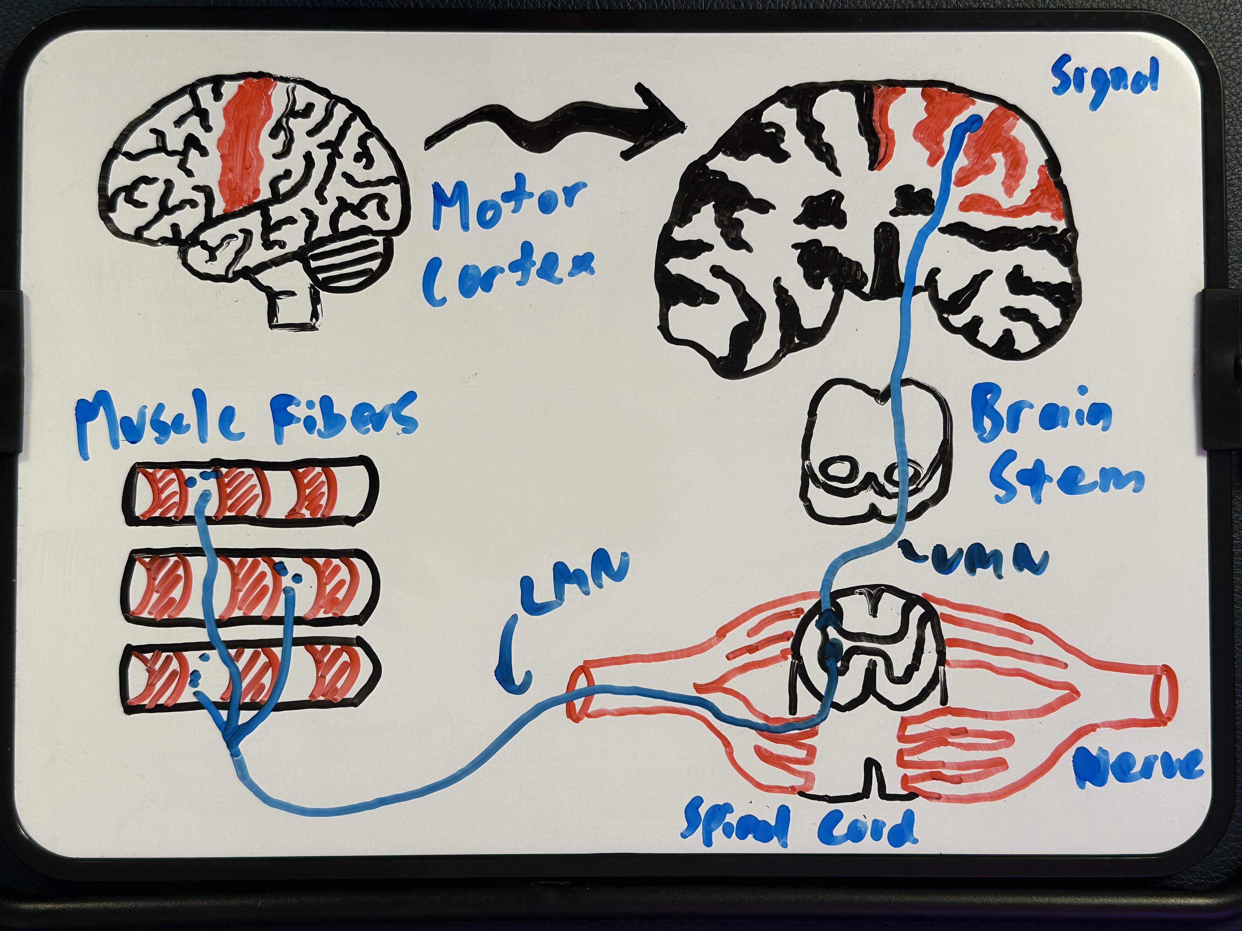
Picture sitting down with your morning coffee, and reaching out to pick up the mug.
That decision to move begins in a section of the cortex called the motor cortex, located within the brain's frontal lobe. In reality, the motor cortex is influenced by many other regions responsible for sensation, planning, and precision — but for now, just know that signal generation within the brain is complex and involves many areas communicating in parallel.
The cortical signal travels down from the motor cortex through the brain and into the spinal cord via a single neuron called an upper motor neuron. This upper motor neuron sends its axon all the way down the spinal cord, where it connects — via a synapse — to a second neuron called a lower motor neuron.
The lower motor neuron exits the spinal cord through a nerve root, becomes part of a peripheral nerve, and travels through your limb until it reaches the target muscle. At the muscle, it forms a specialised synapse called the neuromuscular junction. When the signal arrives, neurotransmitters are released that activate muscle fibres, causing the muscle to contract.
Please keep in mind that this is simplified heavily though. In reality, thousands of signals run through parallel neurons simultaneously, coordinating multiple muscles in real time. But for each individual signal, this two-neuron pathway is the backbone of voluntary movement: one upper motor neuron from brain to spinal cord, and one lower motor neuron from spinal cord to muscle.
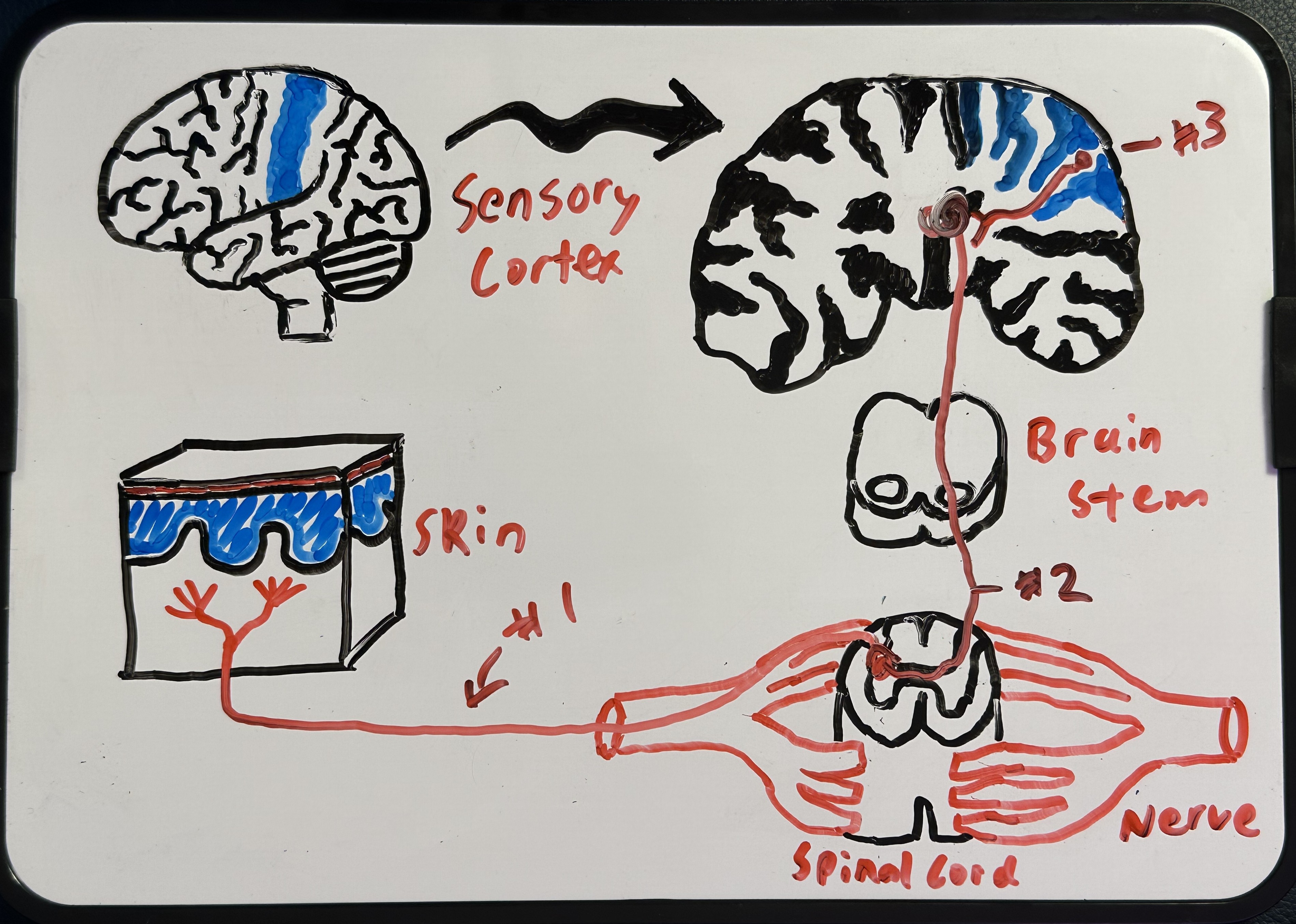
Sensory Input: Body to Brain
Now let's reverse the direction. You've picked up the mug, and it's full of warm coffee!
Specialised sensory receptors in your skin detect that temperature. The receptor — typically the specialised ending of a peripheral sensory nerve — converts that thermal energy into an electrical signal.
That signal travels along a first-order sensory neuron back to the spinal cord. There, it synapses with a second-order sensory neuron, which carries the signal up through the spinal cord to a relay centre deep in the brain called the thalamus.
The thalamus acts like a switchboard, directing sensory information to the appropriate area of the cortex. There, our temperature signal passes from the second-order neuron to a third-order sensory neuron, which carries it to the somatosensory cortex.
At the somatosensory cortex, this signal integrates with many other sensory inputs arriving simultaneously — the pressure and texture of the mug against your skin, the smell of the coffee, how it looks, etc. This sensory integration is what elevates raw sensory data into the conscious experience of holding a warm cup of coffee.
Again, this is simplified. In practice the thalamus distributes signals to numerous brain regions where they are constantly compared, integrated, and filtered. And sometimes, second-order neurons in the spinal cord trigger reflex responses before the brain is even aware.
But in general, the core sensory pathway takes just three neurons: one from receptor to spinal cord, a second from spinal cord to thalamus, and a third from thalamus to cortex.
This constant loop between motor output and sensory input runs every second of your life!
How Things Can Go Wrong
Before wrapping up, it's worth briefly mentioning how different diseases can disrupt this sensory and motor loop, broken down by region.
Damage in the cortex — like in a stroke — can impair signal generation and processing, leading to symptoms like weakness or altered sensations based on which brain region was affected.
Damage to the spinal cord disrupts signal transmission. This can range from patchy regions of weakness and numbness (as in multiple sclerosis) to complete paralysis (as in a severe spinal cord injury).
Damage to peripheral nerves — as in peripheral neuropathy — can again cause numbness or weakness because signals can no longer travel between the spinal cord and the body.
While these seem like the same symptoms, there are important differences in how weakness and numbness present depending on which region is affected; and we'll cover those on a condition-specific basis in future videos/posts.
For now, just remember: while different diseases affect different parts of this pathway, the underlying logic is the same. For clear sensory input and clean motor output, signals must travel from brain to spinal cord to body — and back again — without interruption. If any link in that chain is disrupted, you get symptoms.
Wrapping Up
The nervous system is an extraordinary communication network. It involves billions of interconnected neurons transmitting information via electricity and chemistry. But at its core, it takes just two neurons to send a motor signal from brain to muscle, and just three to relay a sensory signal from skin back to brain!
In future episodes of PK Explains (and corresponding blogposts like this one), we'll break down how specific brain regions specialise in different roles, how the peripheral nervous system divides into further branches, and how individual conditions affect the nervous system in their own unique ways.
For now, you have a foundational understanding of these basic sensory and motor pathways — and we hope that by gaining it, your neurological symptoms have become just a little less mysterious.
References
Lemon, R.N. (2008). Descending pathways in motor control. Annual Review of Neuroscience, 31, 195–218. DOI: 10.1146/annurev.neuro.31.060407.125547
Wang, L.H., Ding, W.Q., & Sun, Y.G. (2022). Spinal ascending pathways for somatosensory information processing. Trends in Neurosciences, 45(8), 594–607. DOI: 10.1016/j.tins.2022.05.005
Stay Informed!
News, updates, and science delivered to your inbox.
We will never sell your information, for any reason.
